A suspension of cells of a spore forming bacterial strain is placed on a microscope slide and spread with a bacteriological ring.
Next follows fixation with mild heating.
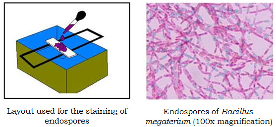
The microscope slide is placed on a grid above a water bath (Τ=100°C). On the surface of the slide is placed an absorbing paper that is soaked through with Malachite green, and remains on the water bath vapors for 5 min. At this phase, the cells and the spores are coloured green. (The bacterial spores are resistant structures and they are not easily soaked through with dyes. For that reason, the staining is performed under heating conditions).
Next, the slide is removed from the water bath and the paper is removed from the slide. The slide is left in room temperature and the excess of dye is removed with deionized water. The water removes the dye from the bacterial cells, but not from the spores.
The sample is covered with safranin for 2 min. During this phase, the cells are colourized red, while the spores remain green.
The excess of safranin is also removed with deionized water.